
Mortality varies depending in part on whether the patient receives timely and appropriate antibiotic therapy. However in milk they are primarily spoilage organisms as the pathogenic species such as Clostridium botulinum and Bacillus anthracis are not associated with milk and relatively few strains of B.

Bacterial Pathogenesis Common Entry Mechanisms Current Biology
1
Molecules Free Full Text Resistance Of Gram Negative Bacteria To Current Antibacterial Agents And Approaches To Resolve It Html
Contact with someone who carries gram negative bacteria.
Gram negative bacteria pdf. Gram stain permits the separation of all bacteria into two large groups those which retain the primary dye gram -positive and those that take the color of the counterstain gram -negative. Gram-negative bacillary sepsis with shock has a mortality rate of 12 to 38 percent. As bactérias Gram-negativa são bactérias que não retêm o corante violeta de genciana durante o recurso ao protocolo de coloração de Gram.
The outer membrane of the gram-negative cell is lost from the cell leaving the peptidoglycan layer exposed. After more than a decade of controversy techniques of electron microscopy were improved to the point in which they finally revealed a clearly layered structure of the Gram-negative cell envelope Fig. THE GRAM-NEGATIVE CELL ENVELOPE.
124 Cubists CB-182804 is a polymyxin B analog currently in phase I trials that shows activity against many MDR gram-negative bacteria and even some colistin resistant strains. Specific to gram-positive bacteria is the presence of teichoic acids in the cell wall. Inhibit growth but gram negative bacteria are protected by their enhanced cell wall and will be able to grow on these plates.
Use Gram staining to see if bacteria are Gram positive or Gram negative. 1Durante o processo de coloração de Gram um corante de contraste normalmente a safranina é adicionado após o violeta de genciana provocando a coloração das bactérias Gram-negativas de rosa ou vermelho. The difference is clear but in simple explanation gram staining is what makes bacteria to be gram positive or negative and this happens because gram positive bacteria have thick peptidoglycan which retains crystal violet staining dye as opposed to gram.
The guava leaves were extracted in four different solvents of increasing polarities hexane. Gram-negative bacterias S-layer is attached directly to the outer membrane. Gram staining is a common technique used to differentiate two large groups of bacteria based on their different cell wall constituents.
MacConkey Agar was first used to isolate the gram-negative bacterium because it inhibits the growth of gram-positive bacteria. Gram-negative bacteria cause infections including pneumonia bloodstream infections wound or surgical site infections and meningitis in healthcare and community settings. Gram in 1884 it remains an important and useful technique to this day.
Gram-positive and gram-negative Bacteria. Gram positive and Gram negative. Pseudomonas spp Binds to bacterial ribosome causing misreading of mRNA resulting in.
To determine the antimicrobial potential of guava Psidium guajava leaf extracts against two gram-negative bacteria Escherichia coli and Salmonella enteritidis and two gram-positive bacteria Staphylococcus aureus and Bacillus cereus which are some of foodborne and spoilage bacteria. Medical devices that pass into the body such as IVs or catheters. A differential stain like that invented by Hans Christian Gram in 1882 will give you more information and allow you to group the stainable bacteria into more groupings.
The mcr gene which is found in gram-negative bacteria encodes a 601-kDa cytoplasmic transmembrane protein 541 amino acids. 331 It was. 1 Glauert and Thornley 1969There are three principal layers in the envelope.
Spore-forming bacteria of the species Bacillus and Clostridium are spoilage organisms that can survive pasteurization but they can also be pathogenic bacteria Doyle et al 2015. Monobactams Aztreonam Gram-positive gram-negative bacteria Inhibition of cell wall synthesis Chromobacterium violaceum Aminoglycosides Streptomycin amikacin gentamicin kanamycin tobramycin neomycin Gram -positive gram negative bacteria esp. Note that the success of the Gram stain relies upon the integrity of the cell wall.
Of all the different classification systems the Gram stain has withstood the test of time. Gram positive bacteria that have been overly heat fixed resulting in destruction of all or parts of their cell wall can appear to be pink Gram negative or have pink areas. Colistin 71 is now used in the treatment of MDR gram-negative pathogens with few other treatment options particularly MDR Pseudomonas Klebsiella and Acinetobacter strains including NDM-1 producers.
Gram-negative bacteria cell wall. The epidemiology microbiology clinical manifestations and treatment of gram-negative bacillary bacteremia will be reviewed here. The Gram stain procedure distinguishes between Gram positive and Gram negative groups by coloring these cells red or violet.
MAC is also differential in identifying gram-negative lactose. Some of these are lipoteichoic acids which have a lipid component in the cell membrane that can assist in anchoring the peptidoglycan. The primary dye is crystal v iolet and the secondary dye is usually either safranin.
The outer membrane OM the peptidoglycan cell wall and the cytoplasmic or. Gram-negative cells have thin layers of peptidoglycan one to three layers deep with a slightly different structure than the peptidoglycan of gram-positive cells Dmitriev 2004With ethanol. The unknown gram-negative bacterium was identified as Escherichia coli.
This is an artifact. All of the biochemical tests worked well except for the indole. They are characterized by their cell envelopes which are composed of a thin peptidoglycan cell wall sandwiched between an inner cytoplasmic cell membrane and a bacterial outer membrane.
The Cell wall of the Gram-Negative Bacteria is very complex as compared to that of Gram-Positive Bacteria. Gram negative bacteria can pass to the body from. The risk increases with the length of the stay.
Thus Gram positives appear deep purple and Gram negatives appear pink. It allows a large proportion of clinically important bacteria to be classified as either Gram positive or negative based on their. Gram staining is a procedure that allows you to divide bacteria into 2 common types.
Gram negative bacterial infections are most common in hospitals. Gram-negative bacteria are bacteria that do not retain the crystal violet stain used in the Gram staining method of bacterial differentiation. Gram-negative bacteria are found in virtually all environments on.
Combined with the major role of the outer membrane of the cell with a layer of peptidoglycan its functional properties are complex and here is a description of the cell wall and its functional parts. Other things that raise the risk. If a culture reveals that a wound is infected susceptibility testing is done to determine which antibiotic will inhibit the growth of the bacteria causing the infection.
Gram stain and bacterial morphology. Selected gram-negative bacteria are becoming resistant to all or nearly all antibiotics meaning that patients with infections from these bacteria might have few or no treatment options. MCR-1 attaches a phosphoethanolamine PEA moiety to the head groups of LPS of lipid A in the gram-negative bacterial membrane 15 which conceals the negatively charged phosphate groups of the bacterial surface where polymyxin E and other polymyxin.
A bacterial wound culture is primarily used along with a Gram stain and other tests to help determine whether a wound is infected and to identify the bacteria causing the infection. If any students are working with a gram positive unknown they pair up with a student with a gram negative unknown since the following methods in this lab were developed for gram. Gram positive rods Gram negative rods Gram postive cocci and Gram negative cocci see images below.
After performing the Gram stain to determine that the unknown was a Gram negative rod the organism was grown on a TSA slant for use in inoculating the rest of the biochemical tests. Gram positive bacteria have an extra thick cellular wall made of a polymer called peptidoglycan that holds a dye stain better than the thinner cell walls of Gram negative bacteria.

Pdf New Antibiotic Molecules Bypassing The Membrane Barrier Of Gram Negative Bacteria Increases The Activity Of Peptide Deformylase Inhibitors
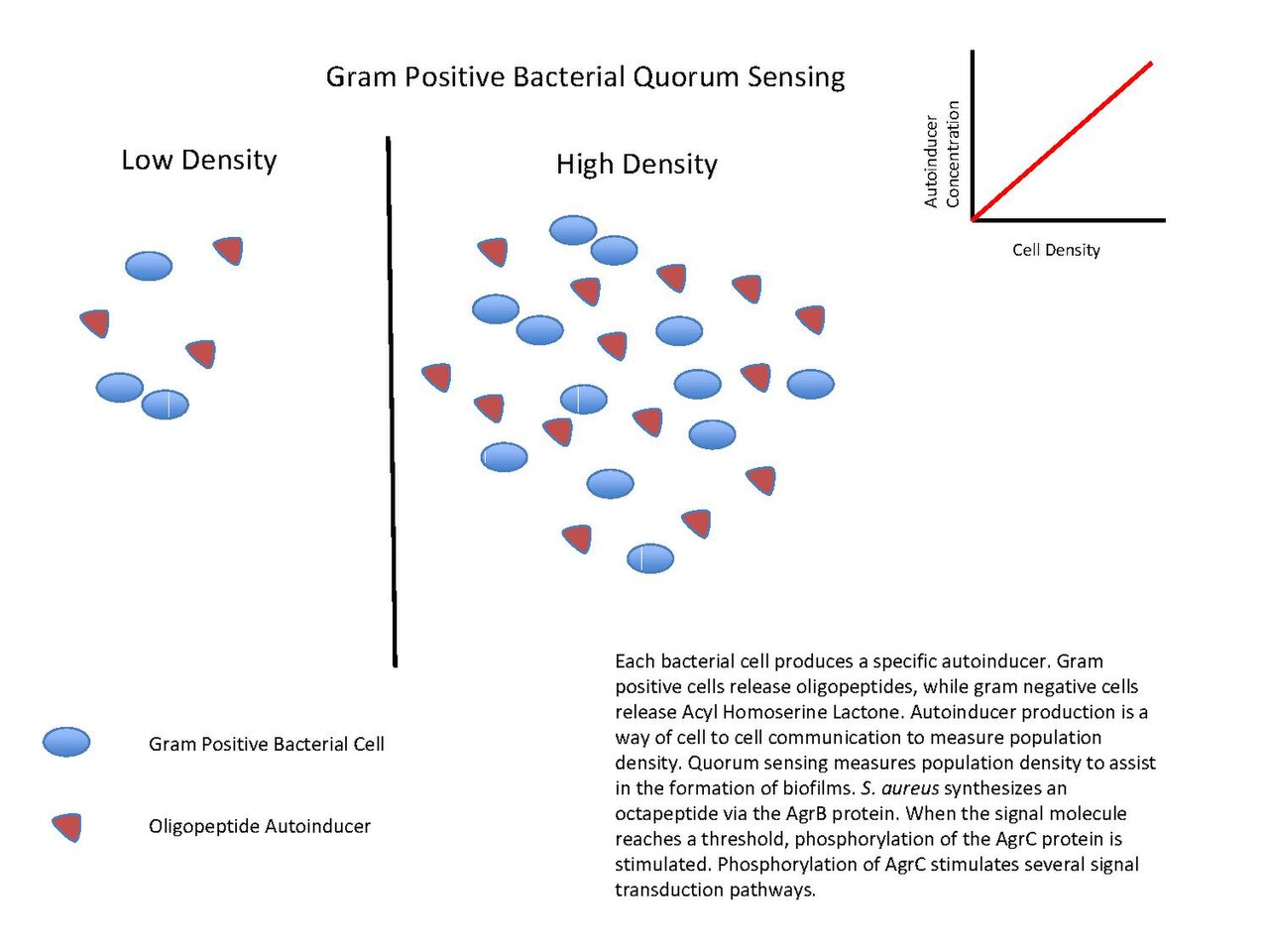
File Gram Positive Bacteria Quorum Sensing Pdf Wikipedia
Pdf Summary Of Biochemical Tests For Some Gram Negative Bacteria

Needs Assessment For Novel Gram Negative Antibiotics In Us Hospitals A Retrospective Cohort Study The Lancet Infectious Diseases
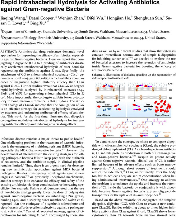
Rapid Intrabacterial Hydrolysis For Activating Antibiotics Against Gram Negative Bacteria Biological And Medicinal Chemistry Chemrxiv Cambridge Open Engage

Antimicrobial Resistance Prediction For Gram Negative Bacteria Via Game Theory Based Feature Evaluation

Are Gram Negative Bacteria Involved In Hla B27 Associated Uveitis British Journal Of Ophthalmology
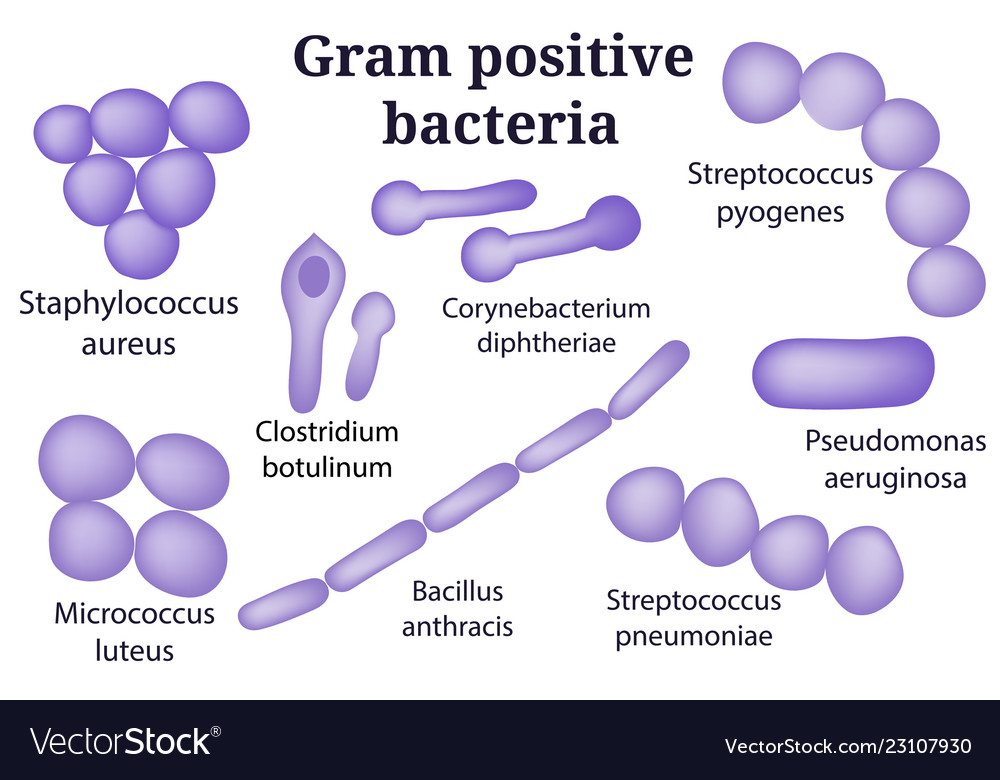
Gram Positive Bacteria Royalty Free Vector Image


